
What is Legg-Calve-Perthes Disease (LCP)?
Legg-Calve-Perthes Disease (LCP) is a disorder of hip joint conformation occurring in both humans and dogs. In dogs, it is most often seen in the miniature and toy breeds between the ages of 4 months to a year.
LCP results when the blood supply to the femoral head is interrupted resulting in avascular necrosis, or the death of the bone cells. Followed by a period of revascularization, the femoral head is subject to remodeling and/or collapse creating an irregular fit in the acetabulum, or socket. This process of bone cells dying and fracturing followed by new bone growth and remodeling of the femoral head and neck, can lead to stiffness and pain.
LCP is believed to be an inherited disease, although the mode of inheritance is not known. Because there is a genetic component, it is recommended that dogs affected with LCP not be used in breeding programs.

Breeds At Risk for Legg-Calve-Perthes
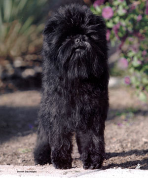
Affenpinscher
Australian Terrier
Bichon Frise
Border Terrier
Boston Terrier
Cairn Terrier
Chihuahua
Cocker Spaniel
Dachshund
Fox Terrier
Jack Russell Terrier
Lakeland Terrier
Manchester Terrier
Miniature Schnauzer
Miniature Pinscher
Pomeranian
Pekingese
Poodle
Pug
Schipperke
Scottish Terrier
Shetland Sheepdog
Silky Terrier
Welsh Terrier
West Highland White Terrier
Yorkshire Terrier
Legg-Calve-Perthes Procedures
In an effort to assist breeders in establishing a control program to limit the prevalence of LCP, the OFA offers a health database specific to LCP. The OFA evaluations and the subsequent database of information will allow breeders to make more informed breeding decisions.
- Owners submit radiographs of their dogs in the standard hip extended ventrodorsal view endorsed by the AVMA.
- Dogs must be a minimum of 12 months of age on the date of the radiograph to be eligible for an LCP number.
- The radiographs may be taken by any veterinarian, but must contain the required dog identification in the film emulsion, exhibit proper positioning, and must be of sufficient quality for the OFA to reach a diagnosis.
- The radiographs along with the completed application and evaluation fee are submitted to the OFA for review.
- At the time of submission, the owner selects whether any abnormal finding may be released in the public domain. All normal results are in the public domain and are available on the OFA website.
- A Board Certified Radiologist reviews the radiograph for evidence of Legg-Calve-Perthes.
- Phenotypically normal dogs are assigned an OFA Legg-Calve-Perthes number.
- Dogs with evidence of LCP are not assigned a number, but the OFA will issue a report stating the findings.
- The OFA submits quarterly reports to the parent club containing the dogs receiving LCP numbers, as well as any overall aggregate statistical data.
The same radiographic image can be used to evaluate the presence of both LCP and hip dysplasia. Evidence of LCP would be detected during an OFA hip dysplasia evaluation and would yield abnormal results. A dog over 12 months of age receiving a normal OFA preliminary report or an OFA hip number is therefore also normal for Legg-Calve-Perthes disease and is automatically eligible to obtain an OFA LCP number.
To receive an OFA LCP number based on a previous hip evaluation, owners should complete the appropriate application and the OFA will assign an LCP number. Evaluation fees will be refunded for dogs determined by the OFA to be affected.
Treatment Options
The degree of clinical severity of LCP varies, and treatment can vary accordingly.
In mild cases, the dog may occasionally resist bearing weight on the affected leg or may exhibit periodic lameness. In these cases, limited activity and treatment with non-steroidal anti-inflammatory drugs (NSAIDs) may be sufficient.
In more severe cases as the pain and discomfort experienced increase, the dog may become totally lame and avoid all use of the affected leg. Furthermore, the leg muscles may begin to atrophy after extended periods of non-use. In severe cases, treatment often resorts to excision of the femoral head and neck. By removing the femoral head and neck, the bone on bone contact that is the source of the pain and discomfort is eliminated. Later, through the healing process and with therapy, a new false joint is formed by muscle and tissue, and the dog may have a complete recovery. Total hip replacement is another treatment option for severe cases now that micro hip replacements have become available.